讲座名称:Stem Cell Imaging-Bench to Bedside-a 15 Year Journey
讲座时间:2019-09-16 14:30:00
讲座地点:南校区G楼118报告厅
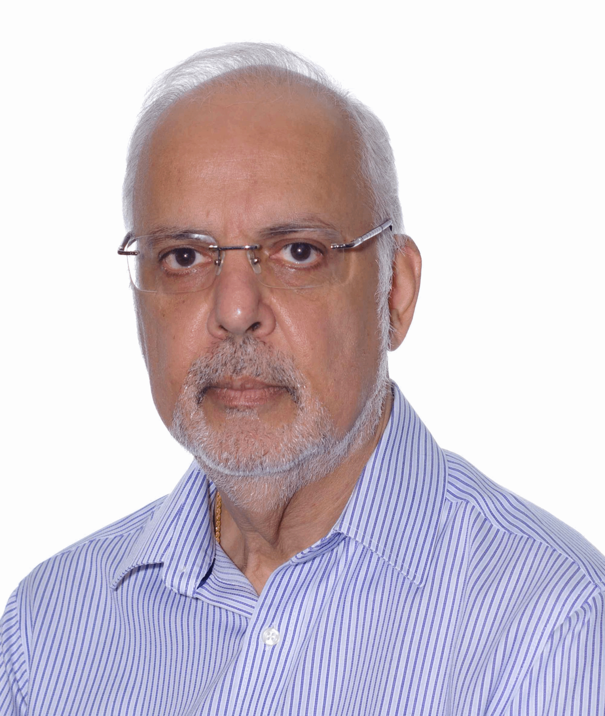
讲座人:Kishore Bhakoo
讲座人介绍:

Kishore Bhakoo教授1978年毕业于坎特伯雷肯特大学医学生物化学专业。他于1983年获得伦敦大学神经病学研究所博士学位,并在路德维希癌症研究所完成博士后培训。 1986年,他在伦敦皇家外科医学院和儿童健康研究所任研究员。1996年,他在牛津大学生物化学系MRC磁共振光谱学系担任研究讲师和职员科学家。2002年在伦敦帝国理工学院MRC临床科学中心担任MRC集团负责人和高级讲师,并成立了干细胞成像小组。2009年,担任新加坡生物成像联盟(SBIC)的转化分子成像组组长。 2011年,担任新成立的翻译影像工业实验室A * STAR的主任。该实验室直接与工业界合作,通过成像技术开发新药。
讲座内容:
Stem cells are currently being evaluated for their therapeutic potential to replace cells in a number of disease or degenerative pathologies. The monitoring of cellular grafts, non-invasively, is an important aspect of the ongoing efficacy and safety assessment of cell-based therapies. Magnetic resonance imaging methods are potentially well suited for such an application, as they produce non-invasive ‘images’ of opaque tissues. For transplanted stem cells to be visualised and tracked by MRI, they need to be tagged so that they are ‘MR visible’. We have developed and implemented a programme of Molecular Imaging in pre-clinical models that is directed towards improving our understanding of in vivo stem cell behaviour in the context of the whole organism.
In order to achieve these goals, we are engineering novel MRI contrast agents and developing specific tagging molecules to deliver efficient amounts of contrast agents into stem cells. The intracellular contrast agents are based on either superparamagnetic nanoparticles, such as polymer-coated iron oxide, or other paramagnetic MR contrast agents.
With its ability to precisely target cell delivery, track cell migration and non-invasively evaluate living subjects over time, this technique will help in the translation and facilitate the clinical realisation and optimisation of stem cell-based therapies. Moreover, it is important that we develop additional multimodal imaging (MRI, PET, SPECT/CT and Optical) methodologies for in vivo monitoring of functional aspects of implanted stem cells.
主办单位:先进材料与纳米科技学院


